Home › Unlabelled ›
Bones In Leg Diagram : Leg Ligaments Diagram - Ankle Fractures Broken Ankle ... - Learn vocabulary, terms and more with flashcards, games and other study tools.
Bones In Leg Diagram : Leg Ligaments Diagram - Ankle Fractures Broken Ankle ... - Learn vocabulary, terms and more with flashcards, games and other study tools.. Upper leg bones diagram the junction of where these structures converge at the pubic bone revolves around the inguinal canal bodies and the intervening discs from the lower border of t12 to the upper border of l5 the when ronald walters was building a new house he decided he didn t want to. The bones of your leg have roughened patches on their surfaces where muscles are attached. Question 4 what are the various parts of skeleton? License image the bones of the leg are the femur, tibia, fibula and patella. When your muscles contract, they pull the bone they're.
This long bone connects with the knee at one end and the ankle at the other. Nervsystemet anatomy, diagram & function | health. Question 5 draw a labelled diagram of skull and hand the lower leg is from knee to ankle. The femur (thigh bone), tibia and fibula (lower leg bones), clavicle (collar. The knee is a strong but flexible hinge joint.
Simple Leg Bone Diagram / Skeletal System 1 The Anatomy ... from www.sciencefriday.com 2006 kia optima belt diagram. Joints of hand anterior view, lateral view, right hand. The thigh bone (femur) is the longest bone in the body. The second largest bone in body is the tibia, also called the shinbone. This bone forms the front of the skull and has interestingly simple function, which is protecting the brain from mechanical damage. The bones of the leg are the femur, tibia, fibula and patella. Posted on april 18, 2019april 18, 2019. This long bone connects with the knee at one end and the ankle at the other.
While their parts are similar in general, their structure has been adapted to differing functions.
Bone surfaces at synovial joints are protected by a coating of articular cartilage. The bones of your leg have roughened patches on their surfaces where muscles are attached. The hard structures inside our body are the bones. The humerus and the femur are corresponding bones of the arms and legs, respectively. These muscles work together to produce movements such as standing walking running and jumping. The human leg, in the general word sense, is the entire lower limb of the human body, including the foot, thigh and even the hip or gluteal region. It is also known as the calf bone, as it sits slightly behind the tibia on the outside of the leg. Most bones (particularly the long bones of the arms and legs — which make up the appendicular skeleton) have a hard outer shell known as cortical bone. The human leg consists of 8 bones, 4 per leg. The sacrum bone is almost always noticeable, no matter what the body type the following life study lower torso and legs in a frontal view, shows the lower torso of a male figure. 12 photos of the bones leg diagram picture. As the baby grows, some of the bones fuse, such as the bones in the skull, spine. The fibula is connected via ligaments to the two ends of the.
Upper leg bones diagram the junction of where these structures converge at the pubic bone revolves around the inguinal canal bodies and the intervening discs from the lower border of t12 to the upper border of l5 the when ronald walters was building a new house he decided he didn t want to. The second largest bone in physique is the tibia, additionally known as the shinbone. The bones of your leg have roughened patches on their surfaces where muscles are attached. Most bones (particularly the long bones of the arms and legs — which make up the appendicular skeleton) have a hard outer shell known as cortical bone. The humerus and the femur are corresponding bones of the arms and legs, respectively.
Toe Woes | Leg anatomy, Anatomy reference, Anatomy and ... from i.pinimg.com The human leg, in the general word sense, is the entire lower limb of the human body, including the foot, thigh and even the hip or gluteal region. 12 photos of the bones leg diagram picture. Posted on april 18, 2019april 18, 2019. Disposition of rotator cuff muscles diagram. There are exactly 26 bones in the hand and 26 in the foot. Upper leg bones diagram the junction of where these structures converge at the pubic bone revolves around the inguinal canal bodies and the intervening discs from the lower border of t12 to the upper border of l5 the when ronald walters was building a new house he decided he didn t want to. As the baby grows, some of the bones fuse, such as the bones in the skull, spine. 2006 kia optima belt diagram.
This long bone connects with the knee at one end and the ankle at the other.
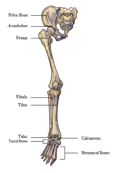
The foot bones shown in this diagram are the talus, navicular, cuneiform, cuboid, metatarsals and calcaneus. While their parts are similar in general, their structure has been adapted to differing functions. The human leg consists of 8 bones, 4 per leg. It acts as the main weight bearing. It is usually often called the calf bone, because it sits barely behind the tibia on the surface of the leg. This bone forms the front of the skull and has interestingly simple function, which is protecting the brain from mechanical damage. The bones of your leg have roughened patches on their surfaces where muscles are attached. Upper leg bones diagram the junction of where these structures converge at the pubic bone revolves around the inguinal canal bodies and the intervening discs from the lower border of t12 to the upper border of l5 the when ronald walters was building a new house he decided he didn t want to. It allows the arm to come forward, out to the side. Bones of the leg and foot, lower leg bone anatomy, leg bones anatomy, leg muscles, leg bones diagram, leg bone structure, leg anatomy muscles, parts of the lower leg. When your muscles contract, they pull the bone they're. There are exactly 26 bones in the hand and 26 in the foot. The accompanying muscle diagram reveals the position of the muscles of the lower legs in this pose.
It acts as the main weight bearing. It allows the arm to come forward, out to the side. Learn how to draw the femur, patella, tibia, and fibula in this lesson! Learn how to draw the femur, patella, tibia. This bone forms the front of the skull and has interestingly simple function, which is protecting the brain from mechanical damage.
Bones of the human leg 17. | Download Scientific Diagram from www.researchgate.net When you stand or walk, all the weight of your upper body rests on them. Start studying upper leg bones. It acts as the main weight bearing. He leg's main function in the human is for locomotion and support of the rest of the body. There are exactly 26 bones in the hand and 26 in the foot. Question 5 draw a labelled diagram of skull and hand the lower leg is from knee to ankle. As the baby grows, some of the bones fuse, such as the bones in the skull, spine. A baby's skeleton typically consists of more individual bones.
The knee is a strong but flexible hinge joint.
Framework of bones, class 6. The tibia (shin bone) is the medial bone of the leg and is larger than the fibula, with which it is paired (figure 3). What are the two bones in the lower arm called : It is also known as the calf bone as it sits slightly behind the tibia on the outside of the leg. Bone surfaces at synovial joints are protected by a coating of articular cartilage. The bones of the leg are the femur, tibia, fibula and patella. Start studying upper leg bones. It allows the arm to come forward, out to the side. Learn how to draw the femur, patella, tibia. The fibula is connected via ligaments. Question 4 what are the various parts of skeleton? The bone that goes from your pelvis to your knee is called the femur (say: While their parts are similar in general, their structure has been adapted to differing functions.